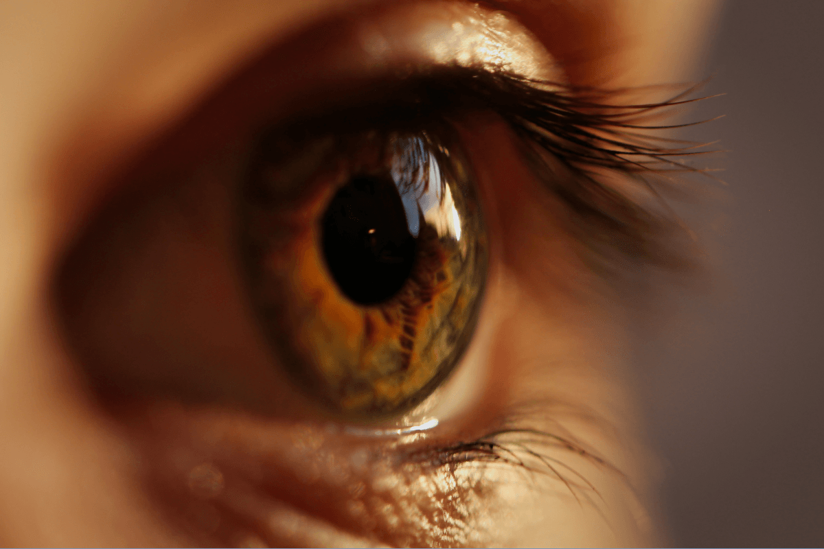
Diabetic retinopathy is an eye complication of diabetes that damages tiny blood vessels in the retina, the light-sensing tissue at the back of the eye. It can develop quietly, so vision may seem normal even while changes are starting. That is why routine dilated eye exams matter for people living with diabetes.
The condition does not mean vision loss is inevitable. Many people protect day-to-day vision through regular screening, careful monitoring, and treatment when retinal swelling or abnormal vessels appear. The best next step depends on your exam findings, symptoms, and overall diabetes health.
Key Takeaways
- Early changes can be silent: Normal vision does not rule out retinal damage.
- Screening finds hidden damage: Dilated exams and imaging can detect small leaks.
- Symptoms deserve context: Blur, floaters, and distortion can have several causes.
- Treatment is individualized: Injections, laser therapy, surgery, or monitoring may be considered.
- Whole-body care helps: Glucose, blood pressure, cholesterol, and kidney health affect risk.
How Diabetic Retinopathy Damages the Retina
Diabetes can harm the retina when long-term high glucose stresses small blood vessels. These vessels may weaken, leak fluid, or close off. The retina can then receive less oxygen, which may trigger growth signals for new fragile vessels.
This process is sometimes described as diabetic retinopathy pathophysiology. In plain language, it means blood vessel injury starts a chain of leakage, swelling, poor blood flow, and repair signals that can become harmful. The macula, the central part of the retina used for reading and face recognition, is especially important.
When fluid collects in or near the macula, clinicians call it diabetic macular edema. This swelling can blur central vision and may occur at different disease stages. It is one reason an eye exam can show risk even when symptoms feel mild.
Diabetic retinopathy causes are not limited to glucose alone. Longer diabetes duration, higher blood pressure, abnormal lipids, kidney disease, pregnancy, and smoking can all influence risk. Rapid changes in glucose control may also affect the eyes, so eye teams often coordinate with diabetes clinicians when treatment plans change.
Why it matters: Eye symptoms are only one part of the picture; imaging can reveal changes before you notice them.
If you are still sorting out diabetes patterns, Type 2 Diabetes Symptoms can help you review common signs to discuss with your care team. For broader context on diabetes types and risk patterns, see Types Of Diabetes.
Warning Signs: What You Might Notice First
The first sign of diabetic retinopathy is often no sign at all. Many people have no pain, redness, or obvious vision change in the early stage. That silence is frustrating, but it is also exactly why screening is emphasized.
When diabetic retinopathy symptoms do appear, they may be subtle at first. Some people notice blur that comes and goes, especially when glucose levels fluctuate. Others report trouble reading, dimmer colors, glare at night, or a smudge near the center of vision.
Floaters can happen when tiny bleeds occur inside the eye. They may look like dots, strings, or cobwebs drifting through vision. A few stable floaters can be common for many reasons, but a sudden shower of new floaters needs prompt advice.
It helps to separate short-lived blur from persistent change. A temporary blur after a large glucose swing may settle, while ongoing distortion or one-sided vision loss should be checked. Straight lines looking wavy, a growing dark patch, or new difficulty seeing faces can suggest macular involvement.
Seek urgent eye care guidance for sudden vision loss, a curtain-like shadow, many new floaters, flashes of light, or severe eye pain. These symptoms can signal problems that need same-day or urgent evaluation, even when diabetes is not the only possible cause.
Stages and Grading: What the Terms Mean
Diabetic retinopathy stages describe how much retinal blood vessel damage an eye clinician sees. Staging helps guide follow-up timing, imaging, and treatment decisions. It is not a judgment about effort or self-management.
The broad categories are nonproliferative diabetic retinopathy and proliferative diabetic retinopathy. Nonproliferative disease means damaged vessels are leaking or closing, but new abnormal vessels have not grown. Proliferative disease means the retina has started growing fragile new vessels because of poor oxygen supply.
Clinicians may further grade nonproliferative disease as mild, moderate, or severe. You may also hear “background diabetic retinopathy,” an older plain-language term often used for earlier nonproliferative changes. The exact grade depends on findings such as microaneurysms, hemorrhages, fatty deposits, and areas of poor blood flow.
| Stage in plain language | What the clinician may see | What you might notice |
|---|---|---|
| Early nonproliferative changes | Tiny vessel bulges or small retinal bleeds | Often no symptoms |
| Moderate nonproliferative changes | More bleeding, leakage, or vessel narrowing | Intermittent blur or mild distortion |
| Severe nonproliferative changes | Larger areas with reduced blood flow | Blur may increase, but vision can still seem good |
| Proliferative changes | New fragile vessels, bleeding, or scar tissue risk | Floaters, sudden blur, dark patches, or vision loss |
Macular status matters as much as the stage. An eye with moderate disease and macular swelling may need more attention than an eye with more widespread changes but no central swelling. Your plan should reflect both the stage and whether the macula is affected.
People often ask whether diabetic retinopathy can be reversed. Some swelling or leakage may improve with treatment and better risk-factor control, but established retinal damage may not fully disappear. The practical goal is to preserve vision, reduce swelling, and lower the chance of worsening damage.
What Happens During Screening and Diagnosis
Diabetic retinopathy screening looks for retinal changes before they threaten daily vision. A standard glasses check is not enough. The visit usually includes dilation or retinal imaging so the clinician can examine the back of the eye.
Most visits begin with visual acuity, eye pressure, and a symptom review. You may be asked about A1C trends, blood pressure, cholesterol, kidney health, pregnancy, and any recent medication changes. Those details help the eye team judge risk and choose follow-up timing.
After dilation, the clinician examines the retina for bleeding, swelling, deposits, abnormal vessels, and scar tissue. Retinal photographs can document small changes. OCT, or optical coherence tomography, creates cross-section images that measure retinal thickness and macular swelling.
Some people also need fluorescein angiography, a dye test that maps leakage and poor blood flow. This test can help when laser treatment is being considered or when the source of vision change is unclear. Your clinician can explain why a test is needed and what the images show.
Quick tip: Ask which eye is affected, whether the macula is swollen, and when follow-up is due.
If eye health is on your prevention list, Healthy Vision Month offers broader reminders about routine exams and daily eye protection. You can also browse Ophthalmology Articles for related eye-health topics.
Treatment Options and How Clinicians Choose Them
Diabetic retinopathy treatment depends on the stage, macular swelling, bleeding risk, and current vision. Some people need monitoring only. Others may need injections, laser therapy, surgery, or a combination over time.
Anti-VEGF injections are commonly used when diabetic macular edema affects central vision or when abnormal vessels need treatment. VEGF is a growth signal that can drive leakage and fragile vessel growth. These injections are given in the eye in a clinical setting after numbing and antiseptic steps.
Steroid treatments may be considered in some cases of macular swelling, but they are not right for everyone. They can affect eye pressure or cataract risk, so clinicians weigh benefits and risks carefully. The choice depends on eye findings, past response, and other health factors.
Laser therapy can help seal or reduce specific leakage patterns. In proliferative diabetic retinopathy, a wider laser pattern may reduce the retina’s oxygen-demand signal and lower bleeding risk. Possible side effects vary by technique and treated area, and your clinician should explain expected vision changes before treatment.
Vitrectomy surgery may be considered when bleeding does not clear, scar tissue pulls on the retina, or a retinal detachment is a concern. Recovery depends on the reason for surgery, the eye’s condition, and any gas bubble or positioning instructions. Your surgeon is the right person to explain restrictions, drops, and follow-up visits.
Medication pages can sometimes help you recognize names discussed in clinic, but they should not guide treatment choices on their own. For example, Eylea and Lucentis Vial are product pages that may provide medication-format context. Decisions about eye injections should come from an ophthalmologist or retina specialist who has examined your eyes.
Readers sometimes ask about the best eye drops for diabetic retinopathy. Drops do not treat the core retinal blood vessel damage in the way injections, laser, or surgery may. Eye drops may still be used for comfort, pressure control, or after procedures when prescribed, but they are not a substitute for retinal care.
Everyday Risk Control That Supports Eye Care
Whole-body diabetes care can reduce ongoing stress on retinal blood vessels. Glucose stability, blood pressure management, lipid care, kidney monitoring, and smoking cessation all matter. These steps support the eye plan, even when eye procedures are also needed.
Blood glucose swings can temporarily blur vision by changing the eye’s fluid balance. That temporary effect is different from retinal bleeding or macular swelling, but symptoms can overlap. A brief log of glucose readings, vision symptoms, and timing may help your clinicians connect patterns.
Blood pressure is especially important because high pressure can worsen leakage from fragile vessels. Cholesterol and triglyceride patterns may also relate to fatty deposits in the retina. If you have kidney disease, pregnancy, anemia, or frequent hypoglycemia, ask your diabetes team how those factors affect eye monitoring.
Some diabetes and weight-management medicines have been studied for eye-related outcomes, but medication decisions need individual review. If you are reading about newer diabetes therapies, Tirzepatide And Retinopathy offers related context, and Wegovy And Vision Loss discusses another vision-safety question. Do not stop or change any prescribed medicine without professional guidance.
For ongoing learning, Diabetes Articles is a browseable collection of diabetes topics. If your clinician mentions eye medication categories, Ophthalmology Options is a browseable product-category list, not a treatment recommendation.
Medical Records, ICD-10 Codes, and Second Opinions
Medical records often use codes to describe diabetic retinopathy in detail. ICD-10 codes may specify the type of diabetes, the eye involved, the stage, and whether macular edema is present. The code supports documentation, billing, and care coordination.
The phrase diabetic retinopathy ICD-10 often appears when people review insurance paperwork or patient portals. In practical terms, the code matters less than the clinical description behind it. Ask what stage you have, whether the macula is swollen, and whether changes were seen in one eye or both.
If you are changing clinics or seeking another opinion, request copies of retinal photos, OCT reports, angiography reports, and visit notes. Images can show change more clearly than a summary line. Keeping dated records can make future comparisons easier.
It is reasonable to ask your clinician to translate terms such as nonproliferative, proliferative, edema, ischemia, hemorrhage, or neovascularization. Understanding these words can make the care plan feel less alarming and more manageable.
Authoritative Sources
The National Eye Institute diabetic retinopathy resource explains causes, symptoms, screening, and treatment options in patient-friendly language.
The American Academy of Ophthalmology overview describes retinal changes, diagnosis, and common treatment approaches.
The CDC diabetes and vision loss page summarizes why regular eye exams matter for people with diabetes.
Recap
Diabetic retinopathy can start silently, but it is not something to ignore. Dilated exams, retinal photos, and OCT scans help find changes before symptoms become obvious. If damage is present, the stage, macular swelling, and bleeding risk guide next steps.
Modern eye care may include monitoring, injections, laser therapy, or surgery when appropriate. Daily diabetes care also supports the retina by reducing stress on small blood vessels. Bring questions to both your eye clinician and diabetes team, especially if symptoms change or your records are unclear.
This content is for informational purposes only and is not a substitute for professional medical advice.