Herpes Treatment focuses on controlling symptoms, shortening outbreaks when possible, and lowering the chance of transmission. There is no cure for herpes simplex virus (HSV) today, but prescription antiviral medicines and practical skin care can make outbreaks more manageable. A clear plan also helps you act early, protect partners, and know when symptoms need urgent medical attention.
Key Takeaways
- Antivirals help manage outbreaks and recurrences.
- Early care may reduce sore duration.
- HSV can spread without visible sores.
- Daily suppression may reduce shedding for some people.
- Eye symptoms, pregnancy, or weak immunity need prompt care.
If you were recently diagnosed, feeling unsettled is normal. HSV is common, and many people learn to manage it with a combination of medicines, safer-sex planning, trigger awareness, and supportive care. The goal is not just fewer sores. It is also less worry, clearer communication, and a plan that fits your health needs.
What Herpes Treatment Can and Cannot Do
Herpes Treatment can reduce symptoms and help prevent future outbreaks, but it does not remove HSV from the body. After infection, HSV stays dormant in nerve cells. It may reactivate later, sometimes after stress, illness, friction, sun exposure, or no clear trigger.
Clinicians usually consider two main approaches. Episodic treatment means antiviral medicine is started when symptoms begin, often during tingling, burning, or early soreness. Suppressive treatment means daily antiviral medicine is used to reduce recurrences and viral shedding. The best approach depends on outbreak frequency, symptom burden, partner risk, pregnancy status, immune health, and personal preferences.
Common prescription antivirals include acyclovir, valacyclovir, and famciclovir. These medicines do not work like antibiotics. Antibiotics treat bacterial infections, while HSV is a virus. For medication-specific reading, see Acyclovir, Valtrex, and Famciclovir as background to discuss with a licensed clinician.
Why it matters: Knowing the limits of treatment prevents false hope and supports realistic planning.
Recognizing HSV Symptoms and Stages
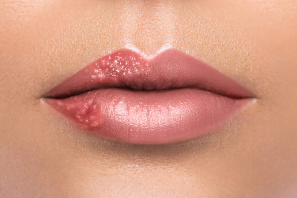
HSV symptoms often follow a recognizable pattern, but not every outbreak looks the same. Some people have painful blisters. Others notice tiny cracks, redness, itching, burning, or soreness that they may mistake for irritation.
A typical outbreak may start with prodrome, which means early warning symptoms before visible sores. Tingling, nerve-like pain, itching, or burning can appear hours or days before blisters. Blisters may then open into ulcers, followed by crusting and healing. First episodes can be more intense and may include fever, swollen glands, body aches, or painful urination. Recurrent outbreaks are often shorter and milder.
Many readers search for herpes photos in different stages because they want reassurance about what they are seeing. Images can help with education, but they cannot confirm HSV. Yeast infections, ingrown hairs, canker sores, shingles, bacterial skin infections, and allergic reactions may look similar. Testing from a fresh sore is often more useful than visual comparison alone.
For a closer look at cold sore patterns, prodrome, and mouth symptoms, read Herpes Symptoms. If sores or pain affect the anal area, Anal Herpes Symptoms explains common signs and when to seek care.
How HSV Spreads, Including Between Outbreaks
HSV spreads through direct skin-to-skin or mucosal contact with an area shedding virus. This can happen during oral, genital, or anal contact. HSV-1 often causes oral herpes, and HSV-2 often causes genital herpes, but either type can affect either location.
Transmission is more likely when sores are present, but HSV can also spread when skin looks normal. This is called asymptomatic shedding. It helps explain why some people acquire HSV from a partner who had no visible symptoms at the time. Condoms and dental dams reduce risk, but they do not cover all exposed skin.
Non-sexual transmission is less common but possible in specific situations, especially with direct contact to active cold sores. Sharing utensils or towels is not considered a major route compared with direct contact, but avoiding contact with sores is still wise. Caregivers should be especially careful around newborns, because neonatal HSV can be serious.
Partner communication works best when it is specific. Discuss where outbreaks occur, what prodrome feels like, when to avoid contact, and whether suppressive therapy makes sense. You can also use broader sexual health education from Understanding STDs to frame safer conversations without blame.
How to Care for Sores and Reduce Discomfort
Early action is the most useful home-care principle. If you have a prescribed episodic plan, ask your clinician how soon to start it after prodrome begins. Do not change dosing or start leftover medicine without medical guidance.
Gentle wound care can reduce irritation while the skin heals. Wash the area with lukewarm water and mild cleanser if needed. Pat dry instead of rubbing. Wear loose, breathable clothing when genital or anal sores are present. Avoid picking, scratching, fragranced products, harsh antiseptics, and sexual contact with active lesions.
For pain, some people use cool compresses or non-prescription pain relievers when those are safe for them. A clinician can advise if pain is severe, urination is difficult, sores are widespread, or symptoms are new. If sores become increasingly red, warm, swollen, or pus-like, a secondary bacterial infection may need evaluation.
People often ask how to heal herpes sores faster. Antiviral treatment started early may shorten an outbreak for some people. Rest, hydration, reduced friction, and avoiding known triggers may also support recovery. These measures help comfort and healing, but they are not a guaranteed way to stop an outbreak immediately.
Quick tip: Keep an outbreak note with symptoms, triggers, and questions for your next visit.
Oral, Genital, Anal, and Eye Herpes Need Different Priorities
The site of HSV symptoms affects your care plan. Oral herpes usually causes cold sores on or around the lips, though sores can occur inside the mouth. Genital herpes may affect the vulva, penis, scrotum, buttocks, thighs, cervix, or rectal area. Anal herpes may cause pain, discharge, bleeding, or sores that are hard to see.
Herpes on lips treatment often includes avoiding direct contact during sores, protecting lips from sun exposure, and discussing antiviral options for frequent or severe cold sores. Some people ask whether oral herpes is curable. It is not curable, but outbreaks can often be managed with a plan.
Eye symptoms need a different level of urgency. HSV near the eye can involve the cornea and may threaten vision. Eye pain, light sensitivity, blurred vision, redness, or discharge during a facial outbreak should prompt urgent medical evaluation. Do not put steroid creams, hydrocortisone, or other topical products near the eye unless a clinician specifically instructs you.
Hydrocortisone cream for herpes is not a standard HSV treatment. Steroids can sometimes worsen infections if used incorrectly. Antiviral cream for herpes may be used in some situations, but topical products generally have limits compared with oral antivirals. If you are comparing topical prescription information, Zovirax Ointment 5% may help you prepare questions for a prescriber.
Daily Suppression, Triggers, and Long-Term Planning
Daily antiviral medication for herpes may be considered when outbreaks are frequent, severe, emotionally distressing, or when reducing transmission risk is a major goal. This decision should be individualized. A clinician may review kidney health, other medicines, pregnancy plans, immune status, and how often symptoms occur.
Trigger tracking can make long-term care more practical. Common triggers include fever, another infection, major stress, poor sleep, menstrual changes, skin friction, and ultraviolet exposure. Not everyone has clear triggers. Still, a simple journal can help you notice patterns and discuss prevention strategies.
Mental health also matters. HSV stigma can cause shame that feels larger than the medical condition itself. Accurate information often helps. HSV is common, manageable for many people, and not a measure of character or cleanliness. If anxiety affects intimacy, sleep, or daily life, consider speaking with a clinician or counselor who understands sexual health.
For broader condition navigation, the Sexual Health collection may help with related prevention and communication topics. The Infectious Disease collection covers other infection-focused education.
Can Herpes Go Away, Become Dangerous, or Be Fatal?
Herpes sores can go away, but the virus remains in the body. After an outbreak heals, HSV becomes dormant. It may reactivate later, which is why recurrences can happen months or years after the first episode.
For most otherwise healthy adults, HSV is not life-threatening. However, context matters. HSV can be dangerous for newborns, people with weakened immune systems, and people with eye or nervous system involvement. Rare complications, such as encephalitis (brain inflammation), require emergency care.
Questions like can herpes kill you or how fast can herpes kill you often come from fear after a new diagnosis. In healthy adults, death from common oral or genital herpes is very uncommon. The situations that need urgent attention include newborn exposure, severe headache with confusion, neck stiffness, seizures, widespread rash, dehydration, eye symptoms, pregnancy with new genital sores, or severe disease in someone immunocompromised.
Leaving herpes untreated for years does not always cause severe harm, especially if outbreaks are rare. But untreated symptoms can still affect comfort, sexual health, and transmission risk. A medical visit can confirm the diagnosis, rule out other infections, and help you decide whether episodic or suppressive therapy is useful.
Similar Conditions and Myths That Cause Confusion
HSV is often confused with other viral skin conditions. Shingles, caused by varicella-zoster virus, can also produce painful blisters, but it behaves differently and often follows a nerve distribution on one side of the body. For a comparison, see Chickenpox vs. Shingles.
Another common myth is that there must be a cure hidden somewhere. Current Herpes Treatment can manage outbreaks and reduce transmission risk, but no approved therapy eradicates HSV from nerve cells. Research continues, including vaccine and cure-related strategies. For a cautious look at the science, read Cure For Herpes.
Over-the-counter products also create confusion. The best over the counter herpes medication depends on what you mean by “best.” Some non-prescription options may ease discomfort, but they do not cure HSV. Prescription antivirals remain the main evidence-based medicines for controlling HSV outbreaks. Ask a clinician before combining creams, especially near mucous membranes, broken skin, or the eyes.
Preparing for a Clinician Visit
A focused visit can turn uncertainty into a plan. Bring a timeline of symptoms, outbreak locations, possible exposures, pregnancy status if relevant, and any past test results. If sores are present, ask whether swab testing is appropriate. Blood tests may help in some cases, but they have limits and should be interpreted carefully.
Useful questions include whether your symptoms fit HSV-1, HSV-2, or another condition; whether episodic or suppressive therapy is reasonable; how to reduce partner risk; and what symptoms require urgent care. If medication access is part of your planning, BorderFreeHealth connects U.S. patients with licensed Canadian partner pharmacies, and prescription details are verified with prescribers when required before pharmacy dispensing.
Herpes Treatment works best when it is practical. The right plan is one you understand, can follow, and can revisit when your relationships, symptoms, or health status change.
Authoritative Sources
For clinical treatment recommendations, review the CDC sexually transmitted infection treatment guidelines.
For public health context and global HSV estimates, see the WHO herpes simplex virus fact sheet.
For patient-facing diagnosis and treatment background, consult the Mayo Clinic genital herpes resource.
This content is for informational purposes only and is not a substitute for professional medical advice.