What is glaucoma? Glaucoma is a group of eye diseases that damage the optic nerve, often gradually and sometimes without obvious early symptoms. It matters because the damage is usually permanent, but early diagnosis and treatment can often slow more vision loss.
Most people hear glaucoma described as an eye pressure problem. Pressure is important, but it is not the whole story. Some people develop glaucoma with high pressure, while others develop normal-tension glaucoma despite readings that are not clearly elevated. The condition also includes several types, and one form, acute angle-closure glaucoma, can cause sudden severe symptoms and needs urgent care.
Key Takeaways
- Glaucoma damages the optic nerve and can slowly reduce vision.
- Early open-angle glaucoma often causes no noticeable symptoms.
- High eye pressure is a major risk factor, but not the only one.
- Treatment aims to protect remaining vision, not restore lost nerve damage.
- Sudden eye pain, halos, nausea, or fast vision changes need urgent care.
What Is Glaucoma and Why Does It Threaten Vision?
Glaucoma is best understood as progressive optic neuropathy (damage to the nerve that carries visual signals from the eye to the brain). When that nerve is injured, the brain receives less visual information, and blind spots can develop. The loss often starts in peripheral vision, so people may not notice a problem until the disease is more advanced.
Inside the eye, a clear fluid called aqueous humor is constantly produced and drained. If drainage is impaired or the optic nerve is especially vulnerable, the pressure inside the eye, called intraocular pressure, may rise enough to harm the nerve. Still, glaucoma is not defined by pressure alone. That is why a normal pressure reading does not fully rule it out, and a high reading does not automatically mean a person already has glaucoma.
Glaucoma stages are usually described as early, moderate, or advanced based on optic nerve and visual field damage, not on how the eye looks from the outside. Early disease may leave central vision intact, while advanced disease can narrow the visual field and affect reading, mobility, and driving.
Why it matters: Lost vision from glaucoma is often permanent, so timing changes the outcome.
Another common question is whether glaucoma can be cured. In most cases, no. Existing optic nerve damage usually cannot be reversed. The goal of care is to protect the vision that remains, lower the chance of progression, and monitor changes over time.
Signs and Symptoms That Need Attention
Many people with the most common form, open-angle glaucoma, have no symptoms early on. That is why regular eye exams matter, especially when risk is higher. By the time noticeable changes appear, some vision may already be lost.
There is no single official 8-sign checklist, but these are common glaucoma warning signs across different types and stages:
- Gradual loss of side vision
- Patchy blind spots
- Tunnel vision in later disease
- Sudden blurred vision
- Halos around lights
- Eye pain or pressure
- Redness in one eye
- Headache, nausea, or vomiting with vision change
Not every person gets the same symptoms. Open-angle disease often progresses quietly. Angle-closure disease can appear suddenly and feel dramatic. Normal-tension glaucoma may also be subtle, with few symptoms until testing finds damage. In infants and young children, excessive tearing, light sensitivity, or a cloudy-looking cornea can be clues to childhood forms.
When symptoms may be an emergency
Severe eye pain, a red eye, halos around lights, nausea, vomiting, and rapid blurry vision can point to acute angle-closure glaucoma. This can raise pressure quickly and threatens sight. Same-day emergency evaluation is important.
Why early signs are easy to miss
Your brain is good at compensating for slow, uneven vision changes. One eye may also cover for the other. That is why saying you can still see fine does not reliably rule out glaucoma.
Common Types of Glaucoma
Glaucoma is not one single condition. The type helps explain how symptoms start, how urgently it needs attention, and how clinicians think about treatment.
| Type | Typical pattern | Why it matters |
|---|---|---|
| Primary open-angle glaucoma | Slow, often silent progression | Most common adult form; routine exams are crucial |
| Angle-closure glaucoma | May be sudden or slowly developing | A sudden attack is an emergency |
| Normal-tension glaucoma | Optic nerve damage despite pressure not clearly high | Shows that pressure is not the whole story |
| Secondary glaucoma | Linked to another eye or health problem | The cause can shape treatment choices |
| Congenital or childhood glaucoma | Begins early in life | Needs prompt specialist evaluation |
Primary open-angle glaucoma is the type many adults hear about first. It usually develops slowly and silently. Angle-closure glaucoma is less common but more dramatic when it occurs suddenly. Secondary glaucoma means another issue, such as inflammation, injury, or medication effects, contributes to the problem.
Some people also hear about chronic angle-closure glaucoma. Unlike an acute attack, it may develop more slowly, but the drainage angle is still crowded or blocked. That is why an eye exam that looks at the angle directly is important.
Congenital glaucoma is rare and involves abnormal eye drainage present at birth or early in childhood. It is a different situation from typical adult glaucoma and needs specialist care.
What Causes It and Who Is at Higher Risk?
The main cause of glaucoma is not one single trigger. In many people, the problem involves reduced drainage of eye fluid, rising intraocular pressure, and gradual optic nerve injury. In others, blood flow, nerve sensitivity, anatomy, or another eye condition may also play a role.
Risk tends to rise with a mix of factors, not just one. Commonly discussed risk factors include:
- Older age
- Family history of glaucoma
- Higher eye pressure
- Long-term corticosteroid use
- Diabetes or other health conditions
- Prior eye injury or eye surgery
- Severe nearsightedness or farsightedness
- Certain racial or ethnic backgrounds
A clinician may also look at corneal thickness, optic nerve appearance, and the shape of the drainage angle. These details help explain why two people with similar pressure readings can have different risk. Risk patterns also vary across populations, so family history and ancestry can influence who needs closer follow-up.
Angle-closure glaucoma has a different mechanical setup. A naturally narrow drainage angle can become blocked more easily, which is why anatomy matters. In susceptible people, certain medicines that widen the pupil or shift eye structures may be discussed as possible triggers, but that risk depends on the person and the type of glaucoma.
Because early open-angle glaucoma can be silent, glaucoma screening usually means regular comprehensive eye exams rather than waiting for symptoms. The best interval depends on age, family history, previous findings, and whether high pressure or optic nerve changes have already been seen.
Ocular Hypertension vs Glaucoma
Ocular hypertension means eye pressure is higher than expected, but there is no clear evidence of optic nerve damage or vision loss yet. Glaucoma means the nerve or visual field already shows damage. The distinction matters because not everyone with ocular hypertension will develop glaucoma, but the risk can be high enough to justify closer monitoring or treatment.
How Glaucoma Is Diagnosed, Treated, and Monitored
Glaucoma is diagnosed with a comprehensive eye exam, not one test alone. Clinicians often combine pressure measurement, a dilated optic nerve exam, visual field testing, and imaging such as optical coherence tomography, or OCT, which maps the nerve fiber layer. Gonioscopy, a close look at the drainage angle, helps identify whether the angle is open or narrow.
Glaucoma treatment usually aims to lower eye pressure and slow progression. Depending on the type and severity, care may include prescription eye drops, laser procedures, surgery, or a combination. The right plan depends on how advanced the disease is, how fast it seems to be changing, and how well a person can stay consistent with treatment.
What treatment is trying to do
Eye drops may reduce fluid production or improve drainage. Laser treatment may help fluid leave the eye more effectively or reduce fluid made inside the eye, depending on the situation. Surgery is considered when the nerve is at risk, pressure goals are not met, or the disease continues to progress despite other measures.
Follow-up matters because glaucoma stages are defined by the amount of optic nerve and visual field damage, and those changes can be gradual. A person may feel stable even while tests show progression. Many people retain useful vision for years when the condition is found early and followed closely, but that usually requires ongoing monitoring rather than a one-time fix.
Daily treatment can be hard to sustain, especially when symptoms are absent. Dryness, redness, cost, hand dexterity, and confusion about timing can all get in the way. Raising those barriers early can help the care team adjust the plan before adherence slips.
Quick tip: Bring a current medication list, including steroids and eye drops, to every eye visit.
- Ask what type you have
- Ask whether vision loss is present
- Ask which tests will track change
- Ask how often follow-up matters
- Ask what side effects to report
- Ask which symptoms need urgent review
When required, the dispensing pharmacy may confirm prescription details with the original prescriber.
Living With Glaucoma and Related Questions
Living with glaucoma often means building habits around drops, appointments, and routine testing. That can feel frustrating because glaucoma may not cause daily symptoms, yet the condition still needs attention. A refill plan, reminder system, and clear understanding of your follow-up schedule can make long-term care easier to sustain.
People also ask whether certain foods trigger glaucoma or whether diet can fix it. There is no single food known to cause glaucoma in everyone, and diet is not a substitute for treatment. A generally healthy eating pattern supports overall health, but glaucoma management still depends on monitoring and pressure control. If supplements, caffeine, or other habits are a concern, they are best discussed in the context of the specific glaucoma type and the rest of a person’s health history.
Daily life considerations can include lighting, contrast, fall prevention, and asking whether driving is still safe if visual field loss has progressed. Some people also benefit from low-vision services, especially when glaucoma is advanced. These supports do not treat the disease, but they can help preserve independence.
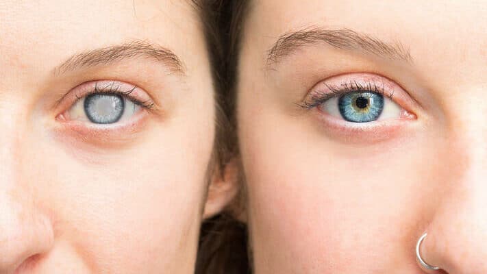
Glaucoma vs Cataracts
These conditions both affect vision, but they are not the same. Cataracts cloud the eye’s natural lens and often cause blurry or dim vision that can usually be treated with surgery. Glaucoma damages the optic nerve. The vision loss from glaucoma is often permanent, which is why early detection matters so much.
For broader eye-health reading, visit our Ophthalmology hub. If you are reviewing prescribed eye therapies, the Ophthalmology Products section groups eye-care products by category.
BorderFreeHealth works with licensed Canadian partner pharmacies for eligible U.S. patients.
Further reading can help, but glaucoma care still depends on individual findings such as eye pressure, optic nerve changes, visual field results, and symptom pattern. If the diagnosis is new, the most useful next step is often understanding the type of glaucoma involved, the goal of treatment, and which warning symptoms should never wait.
Authoritative Sources
- National Eye Institute overview of glaucoma
- American Academy of Ophthalmology patient information
- MedlinePlus summary of glaucoma basics
Glaucoma can be quiet, but it is not harmless. Knowing the type, the warning signs, and the purpose of treatment can make follow-up feel less overwhelming.
This content is for informational purposes only and is not a substitute for professional medical advice.