Key Takeaways
- Often silent early: Vision may stay normal at first.
- Screening matters: Dilated exams can catch changes sooner.
- Many options exist: Injections, lasers, and surgery may help.
- Whole-body control helps: Glucose, blood pressure, and lipids matter.
Eye changes can feel frustrating, especially when vision seems “fine.” Diabetic retinopathy is one reason clinicians stress routine eye checks. It can develop slowly, without pain or obvious symptoms.
Below is a clear, step-by-step look at what happens in the retina. You will also learn what to expect at exams, and how treatments are chosen. The goal is to help you feel informed, not overwhelmed.
If anything here sounds like your experience, an eye clinician can help sort out next steps. Many people protect vision by catching changes early and monitoring them closely.
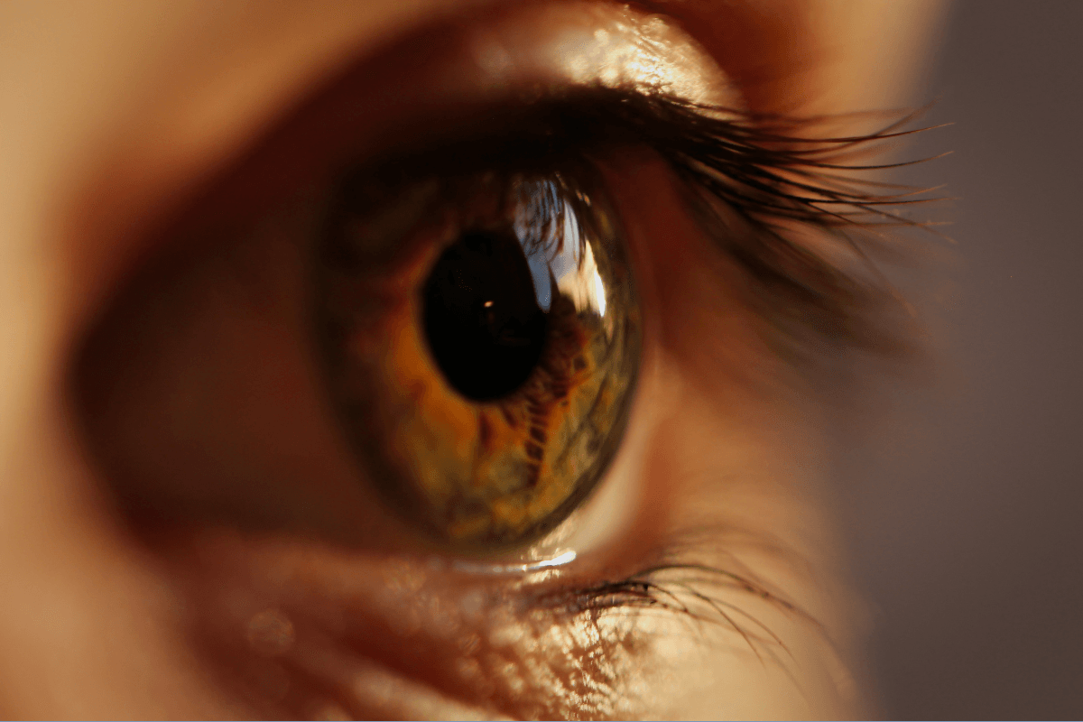
Diabetic Retinopathy: What Changes Inside the Eye
Your retina is a thin layer of light-sensing tissue at the back of the eye. It needs steady blood flow and healthy small vessels. Over time, higher glucose levels can stress those tiny vessels.
When vessel walls weaken, small leaks may develop. Fluid and fats can seep into retinal tissue. In other areas, reduced blood flow can leave the retina short on oxygen.
Clinicians often describe this as a chain reaction. The steps can vary by person, but the core pattern is similar. It helps explain why regular exams matter, even with good vision.
Vessel injury: Small capillaries become fragile and narrow.
Leakage and swelling: Fluid can blur central vision if the macula swells.
Oxygen signal: The retina releases growth signals under low oxygen.
Abnormal vessels: New vessels may grow, but they are weak.
Those growth signals are one reason certain injections can help. They aim to reduce abnormal vessel growth and swelling. For a plain-language overview from a federal eye health source, read the National Eye Institute page with background and screening advice.
Early Warning Signs and Vision Changes
Not everyone notices symptoms early, and that is common. Still, certain changes deserve attention. Keeping track of new visual changes can help you describe them clearly at your visit.
Some people report blur that comes and goes, especially with fluctuating glucose. Others notice trouble reading, dimmer colors, or more glare at night. Dark specks (floaters) may appear when tiny bleeds happen inside the eye.
It can help to separate daily fluctuations from persistent changes. Brief blur after a large glucose swing may settle. Ongoing distortion, straight lines looking wavy, or a growing smudge needs prompt evaluation.
When people search for diabetic retinopathy symptoms, they often want to know what is “normal” versus urgent. A useful rule of thumb is to seek care sooner when symptoms are new, worsening, or one-sided.
Note: Sudden vision loss, a curtain-like shadow, or many new floaters can be urgent. An eye clinician can advise the right timing based on your symptoms.
If you are also seeing signs of high glucose, it may help to review patterns. For a practical refresher on warning signs and common triggers, read Hyperglycemia Signs And Symptoms for symptom context and discussion points.
Stages of Retinal Damage From Diabetes
Clinicians often describe disease progression in stages. The purpose is not to label you. It is to guide monitoring intervals and match treatment to risk.
Early stages usually involve small vessel changes and tiny bleeds. Later stages can include areas of poor blood flow and more widespread leakage. The most advanced stage involves new abnormal vessel growth, which raises bleeding and scarring risks.
Even within a stage, people can have different symptoms. Some have excellent vision despite clear retinal findings. Others have blur mainly from macular swelling, which can happen at more than one stage.
Here is a simplified table that many clinics use to explain progression. Your clinician may use slightly different terms based on imaging and exam findings.
| Stage (plain language) | What an eye clinician may see | What you might notice |
|---|---|---|
| Early changes | Tiny microbleeds, mild leakage | Often no symptoms |
| Moderate changes | More hemorrhages, vessel narrowing | Intermittent blur, mild distortion |
| Severe changes | Large areas with poor blood flow | Blur may increase, night vision worsens |
| Advanced growth stage | New fragile vessels, bleeding risk | Floaters, sudden blur, dark patches |
One search term you may see is diabetic retinopathy stages. In practice, staging is combined with macular status, especially whether swelling affects central vision. That combination often drives treatment choices.
If you want a broader look at how vision changes across the lifespan, including other common conditions, Vision Changes With Age offers helpful comparison points and exam reminders.
What an Eye Exam Usually Includes
A diabetes-focused eye visit is not only a vision test. It is also a health check of the retina and optic nerve. Many key findings are only visible after pupil dilation.
Visits often start with vision, eye pressure, and a review of symptoms. You may be asked about recent glucose changes, blood pressure, pregnancy, or kidney health. These details can affect risk and follow-up timing.
After dilation, the clinician examines the retina for bleeding, swelling, and vessel changes. Photos and scans add detail that the naked eye cannot capture. These tools also help track change over time.
Imaging tests: photos, OCT, and angiography
Retinal photos document small hemorrhages and fatty deposits. OCT (optical coherence tomography) is a scan that shows cross-sections of the retina. It can measure swelling in and near the macula with high precision. Some clinics also use fluorescein angiography, a dye test that maps blood flow and leakage. The best test mix depends on your findings and symptoms.
Ask for a simple explanation of the images you see. Many clinics will point out leakage, swelling, or “non-perfused” areas. Understanding those terms can make future visits feel less confusing.
For general standards around eye exams and diabetes care, the American Diabetes Association has a patient-friendly overview you can review before appointments.
Treatments That Can Help Protect Vision
Treatment decisions depend on location and severity of retinal changes. Your symptoms matter, but imaging matters too. Some people need careful monitoring only, while others benefit from earlier intervention.
The phrase diabetic retinopathy treatment covers several approaches. The most common options aim to reduce swelling, prevent bleeding, or slow abnormal vessel growth. Your eye clinician may also coordinate with your primary diabetes team, since whole-body control supports eye outcomes.
Anti-VEGF injections are commonly used when swelling affects central vision or when abnormal vessels are present. They are delivered in-office with numbing steps to reduce discomfort. If you want to understand medication types and handling details, see Eylea for an example product page with formulation notes and storage basics.
Laser therapy may be used to reduce the risk of bleeding in advanced disease. It can also be used in targeted ways for specific leakage patterns. People often worry about pain, but clinics typically use numbing measures and clear aftercare instructions.
Surgery (often vitrectomy) may be considered when bleeding does not clear, or when scar tissue threatens the retina. Recovery looks different for each person and depends on the problem being addressed. Your surgeon can explain activity limits, eye drops, and follow-up timing in plain terms.
If you are comparing anti-VEGF options based on dosing schedules or device type, Lucentis Vial is another example product page where you can review the formulation format and packaging.
For more detailed clinical explanations of treatment categories and why they are chosen, the American Academy of Ophthalmology provides a clear overview written for patients.
Day-to-Day Diabetes Care That Supports Eye Health
Eye care works best when it fits into your overall health plan. Blood glucose, blood pressure, and cholesterol all affect small blood vessels. Even modest improvements can reduce ongoing stress on the retina.
It can also help to avoid big swings in glucose when possible. Large fluctuations may temporarily blur vision and make patterns harder to interpret. If you are unsure whether symptoms track with glucose, keeping a brief log can support a better conversation at your next visit.
Some life stages and conditions can raise risk, such as pregnancy, kidney disease, and anemia. Sleep, mental health, and access to healthy food also matter, and these are not “willpower” issues. If care feels hard to manage, it is reasonable to ask about support resources and simpler plans.
Because diabetes affects many organs, learning across topics can be useful. For example, Diabetic Kidney Disease explains microvascular (small-vessel) strain in another organ system. Diabetic Neuropathy covers nerve effects that sometimes travel with similar risk factors.
If you are newer to diabetes or still sorting out symptoms, Type 2 Diabetes Symptoms can help you separate common signs from less typical ones. For broader browsing on eye and diabetes topics, Ophthalmology Articles and Diabetes Articles can help you find related explainers.
Documentation and Coding in Medical Records
Medical notes can look intimidating, especially when they include codes and abbreviations. Those details are mainly used for documentation, billing, and care coordination. They can also help ensure the right follow-up and testing are approved.
You may see ICD-10 codes that specify the eye involved, the stage, and whether macular swelling is present. Some codes are very specific, while others are “unspecified” when details are still being clarified. If something looks off, it is appropriate to ask the clinic to explain what each part means.
The term diabetic retinopathy icd-10 may show up when people review their charts or insurance paperwork. In everyday care, what matters most is the clinical description behind the code. That includes imaging results, your vision, and the plan for monitoring or treatment.
If you are collecting records between clinics, ask for copies of your retinal photos and OCT reports. Images can show change more clearly than text alone. Keeping a dated folder on your phone or computer can make second opinions easier.
When you want to compare eye medication categories beyond this article, Ophthalmology Options is a browsable list that can help you recognize common drug classes. It is not a substitute for clinician advice, but it can make treatment conversations easier to follow.
Recap
Diabetes can affect the retina slowly and quietly, especially early on. Regular dilated exams and imaging help catch changes before they threaten daily vision. If problems are found, monitoring and modern treatments can often reduce risk of worsening damage.
Supportive diabetes care also matters, including glucose stability and blood pressure control. If you are unsure what your notes mean or what comes next, bring questions to your eye clinician and diabetes team.
This content is for informational purposes only and is not a substitute for professional medical advice for your personal situation.